Optical Microscopy

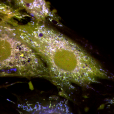
Mitochondria
INFO Service
CAST's optical microscopy service offers the opportunity to evaluate a variety of samples, from cell monolayers to microorganisms as well as tissues sections of the brain and other organs. The service provides a variety of optical imaging systems, including confocal laser scanning systems (LSCM), Multi-Photon (MP) and open-field microscopy. A conventional fluorescence microscopy system is also available for high-throughput screening. These technologies allow wide applications in the field of bio-imaging, thereby providing the investigation of cellular and subcellular structure and function. These methods allow the investigation of localizations and co-localizations of biological molecules at the cellular and subcellular levels. Advanced microscopy to investigate in cells and biological tissues as well as at the single-cell level, the morphology of the cytoskeleton and organelles (i.e., lysosomes, endosomes, mitochondria), their vesicular trafficking and cytotoxicity-related events. Furthermore, protocols for the study of dynamic intracellular events in live cells allow the assessment of changes in the level of intracellular calcium and other second messengers, the analysis of the production of reactive oxygen species, the investigation of mitochondrial functioning.

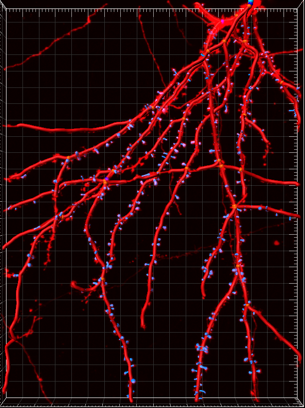
Dendrites of cultured neurons
INFO Access
The service is set to provide basic and advanced training for the CAST researchers on the correct use of microscopes as well as on how to perform experiments independently. For this purpose, researchers are required to attend an introductory course held by the Confocal Microscopy staff before being allowed to use any tool.

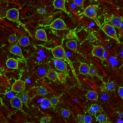
Neuronal staining in hippocal slice
Zeiss LSM 5 PASCAL Confocal Microscope
The Zeiss LSM 5 is a confocal fluorescence microscopy system that provides high-resolution images and three-dimensional reconstructions by means of optical sectioning. The system is based on a Zeiss Axiovert 200 microscope equipped with 10X Plan-Neo / 0.3 NA, 20X Plan-Apo / 0.75 NA, 40X Plan-Neo / 1.4 NA Oil 63X Plan-Apo / 1.3 NA Oil and 100X Plan-Apo / optics 1.4 NA equipped with three laser excitation sources: Argon 458/477/488 / 514nm at 30mW, HeNe 543nm at 1mW and finally HeNe 633nm at 3mW. The laser excitation power is modulated by means of an interference filter and selected with the following set of filters: HFT 405/488/543/633, HFT 405/488/543, FT 458/514, HFT 458/543, HFT 458 and HFT 488. The acquisition system uses two conventional photomultipliers (PMT, with 8 or 12 bit sampling for 256 or 4096 gray levels respectively). The scanning head is based on galvanometric mirrors and the emission, addressed to the PMTs, can be selected by combining the following dichroic and emission filters NFT 490, NFT 545, NFT KP 545 NFT 635 vis, LP 475, LP 505, BP 470-500, BP 475-525, BP 505-530, BP 505-530, BP 505-550, BP 530-600, BP 560-615. The microscopy system is implemented with a Zeiss incubation system that allows time-lapse and 3D acquisitions, and 4D observation of live biological samples by employing a thermostat control and CO2 control (TempModule S1, CO2 Module S1, Control Sensor T S1 with the inserts for the Heating Insert P S1 microscope, Heating Insert M06 S1). In addition, the microscope is equipped with an AxioCam MRC 5 camera and Axiovision software to allow the acquisition of images in conventional fluorescence or bright field settings. ESFRI classification: Health and Food Domain; Acquisition year: 2003READ MORE
Zeiss LSM 7 MP NLO Multiphoton Microscope
Multi-photonic fluorescence microscopy system for live imaging of cell and tissues that employs a Zeiss Axio Examiner microscope equipped with W-Plan-Apo 10X / 0.3 NA, W-Plan-Apo 20X / 1.0 NA VIS-IR DIC, W-Plan-Apo 40X / 1.0 NA DIC, W-Plan-Apo 63X / 1.0 NA VIS-IR DIC. The acquisition system has an NDD module (BP 500-550 / SP485) and a GaAsP-NDD reflection detector, a BiG module for transmitted light (BP 380-430 / 470-520; BP 420-475 / 500-550 ; BP 500-550 / 575-620; BP 525-570 / LP650) as well as a TD detector. The excitation source uses an IR mode-locked Ti: S Laser Coheren Camaleon Vision II of 3W of power with emission modulable from 680 to 1080 nm. ESFRI classification: Health and Food Domain; Acquisition year: 2011READ MORE
Zeiss LSM 800 URGB Confocal Microscope
The technology is based on the Zeiss AXIO OBSERVER.Z1 microscope equipped with Plan-Neo 20X / 0.50, Plan-Neo 40X / 1.30 OIL DIC optics and Plan-Apo 63X / 1.40 OIL DIC and motorized stage. A confocal fluorescence microscopy system that provides high-resolution images and three-dimensional reconstruction by means of optical sectioning. The system is equipped with 4 solid-state laser excitation sources 405nm (5mW), 488nm (10mW), 561nm (10mW), 640nm (5mW). The detectors within the scanning head use the Variable Secondary Dichroic system which allows the selection of the spectral range sent to the PMT and makes spectral imaging possible. The system is equipped with two GaAsP-PMT modules and a third channel based on the Airyscan system (32 PMT / 0.2 Airy unit functional matrix). Finally, the microscopy system is equipped with a detector for transmitted light and an ESID-type lighting system with interference contrast. ESFRI classification: Health and Food Domain; Acquisition year: 2017READ MORE
Olympus ScanR High-Throughput Screening Station fluorescence microscope
Conventional fluorescence microscopy system based on an IX81 Olympus microscope equipped with a Märzhäuser SCAN IM motorized stage and an automatic plate loading system using a Hamilton robotic arm. The lighting system is based on the CellR module with 75W Xenon lamp and 100W transmitted light with DIC. The image acquisition system is based on a Hamamatsu ORCA R2 (CCD) camera and is equipped with WD 0.1- 0.2mm lenses (magnifications: 10x, 20x, 40x, 60x). The following excitation filters are available in the kit: DAPI ( 350/50); CFP (430/25); GFP (470/22); FITC (492/18); YFP (500/20); mRFP (556/20); TxRed (572/23); Cy5 (640/30). The emission filters are: DAPI / FITC / TRITC (U-61002bs, U-61002m), CFP / YFP GFP / RFP (U-51019x, U-51019bs, U-51019m), HqFITC (Dichroic 505LP, emission 535/50 ), Cy5 (Dichroic 660LP, emission 700 / 75m). The system is managed by two different modules: the ScanR Acquisition and Analysis software. The analysis is possible both online and offline and based on the multicore types that allow particle analysis and recognition, extraction of timelapse or static parameters and their calculation. The system allows cytometric expression of data (dot plot) and classification. ESFRI classification: Health and Food Domain; Acquisition year: 2016READ MORE
Granzotto A, Bomba M, Castelli V, Navarra R, Massetti N, d'Aurora M, Onofrj M, Cicalini I, Del Boccio P, Gatta V, Cimini A, Piomelli D, Sensi SL. Inhibition of de novo ceramide biosynthesis affects aging phenotype in an in vitro model of neuronal senescence. Aging (Albany NY). 2019 Aug 29;11(16):6336-6357. DOI: 10.18632/aging.102191
Puca V, Traini T, Guarnieri S, Carradori S, Sisto F, Macchione N, Muraro R, Mincione G, Grande R. The Antibiofilm Effect of a Medical Device Containing TIAB on Microorganisms Associated with Surgical Site Infection. Molecules. 2019 Jun19;24(12). DOI: 10.3390/molecules24122280
Frazzini V, Granzotto A, Bomba M, Massetti N, Castelli V, d'Aurora M, Punzi M, Iorio M, Mosca A, Delli Pizzi S, Gatta V, Cimini A, Sensi SL. The pharmacological perturbation of brain zinc impairs BDNF-related signaling and the cognitive performances of young mice. Sci Rep. 2018 Jun 27;8(1):9768. DOI: 10.1038/s41598-018-28083-9
Bomba M, Granzotto A, Castelli V, Massetti N, Silvestri E, Canzoniero LMT, Cimini A, Sensi SL. Exenatide exerts cognitive effects by modulating the BDNF-TrkB neurotrophic axis in adult mice. Neurobiol Aging. 2018 Apr;64:33-43. DOI: 10.1016/j.neurobiolaging.2017.12.009
Verginelli F, Perconti S, Vespa S, Schiavi F, Prasad SC, Lanuti P, Cama A, Tramontana L, Esposito DL, Guarnieri S, Sheu A, Pantalone MR, Florio R, Morgano A, Rossi C, Bologna G, Marchisio M, D'Argenio A, Taschin E, Visone R, Opocher G, Veronese A, Paties CT, Rajasekhar VK, Söderberg-Nauclér C, Sanna M, Lotti LV, Mariani-Costantini R. Paragangliomas arise through an autonomous vasculo-angio-neurogenic program inhibited by imatinib. Acta Neuropathol. 2018 May;135(5):779-798. DOI: 10.1007/s00401-017-1799-2
Capasso L, D'Anastasio R, Guarnieri S, Viciano J, Mariggiò M. Bone natural autofluorescence and confocal laser scanning microscopy: Preliminary results of a novel useful tool to distinguish between forensic and ancient human skeletal remains. Forensic Sci Int. 2017 Mar;272:87-96. DOI: 10.1016/j.forsciint.2017.01.017
Capone V, Clemente E, Restelli E, Di Campli A, Sperduti S, Ornaghi F, Pietrangelo L, Protasi F, Chiesa R, Sallese M. PERK inhibition attenuates the abnormalities of the secretory pathway and the increased apoptotic rate induced by SIL1 knockdown in HeLa cells. Biochim Biophys Acta Mol Basis Dis. 2018 Oct;1864(10):3164-3180. DOI: 10.1016/j.bbadis.2018.07.003
Cataldi A, Gallorini M, Di Giulio M, Guarnieri S, Mariggiò MA, Traini T, Di Pietro R, Cellini L, Marsich E, Sancilio S. Adhesion of human gingival fibroblasts/Streptococcus mitis co-culture on the nanocomposite system Chitlac-nAg. J Mater Sci Mater Med. 2016 May;27(5):88. DOI: 10.1007/s10856-016-5701-x
Guerra E, Trerotola M, Tripaldi R, Aloisi AL, Simeone P, Sacchetti A, Relli V, D'Amore A, La Sorda R, Lattanzio R, Piantelli M, Alberti S. Trop-2 Induces Tumor Growth Through AKT and Determines Sensitivity to AKT Inhibitors. Clin Cancer Res. 2016 Aug 15;22(16):4197-205. DOI: 10.1158/1078-0432.CCR-15-1701
Pompilio A, De Nicola S, Crocetta V, Guarnieri S, Savini V, Carretto E, Di Bonaventura G. New insights in Staphylococcus pseudintermedius pathogenicity: antibiotic-resistant biofilm formation by a human wound-associated strain. BMC Microbiol. 2015 May 21;15:109. DOI: 10.1186/s12866-015-0449-x
Caprara GA, Perni S, Morabito C, Mariggiò MA, Guarnieri S. Specific association of growth-associated protein 43 with calcium release units in skeletal muscles of lower vertebrates. Eur J Histochem. 2014 Dec 5;58(4):2453 DOI: 10.4081/ejh.2014.2453
Guarnieri S, Morabito C, Paolini C, Boncompagni S, Pilla R, Fanò-Illic G, Mariggiò MA. Growth associated protein 43 is expressed in skeletal muscle fibers and is localized in proximity of mitochondria and calcium release units. PLoS One. 2013;8(1):e53267. DOI: 10.1371/journal.pone.0053267
Grande R, Di Giulio M, Bessa LJ, Di Campli E, Baffoni M, Guarnieri S, Cellini L. Extracellular DNA in Helicobacter pylori biofilm: a backstairs rumour. J Appl Microbiol. 2011 Feb;110(2):490-8. DOI: 10.1111/j.1365-2672.2010.04911.x
Pompilio A, Crocetta V, Confalone P, Nicoletti M, Petrucca A, Guarnieri S, Fiscarelli E, Savini V, Piccolomini R, Di Bonaventura G. Adhesion to and biofilm formation on IB3-1 bronchial cells by Stenotrophomonas maltophilia isolates from cystic fibrosis patients. BMC Microbiol. 2010 Apr 7;10:102. DOI: 10.1186/1471-2180-10-102
Salvatorelli E, Guarnieri S, Menna P, Liberi G, Calafiore AM, Mariggiò MA, Mordente A, Gianni L, Minotti G. Defective one- or two-electron reduction of the anticancer anthracycline epirubicin in human heart. Relative importance of vesicular sequestration and impaired efficiency of electron addition. J Biol Chem. 2006 Apr 21;281(16):10990-1001. DOI: 10.1074/jbc.M508343200
