Scanning Electron Microscopy and EDXA
INFO Service
The scanning electron microscope (SEM) and microanalysis (EDS) facility at CAST offers services for academic researchers and external private/company users. The SEM-EDS Phenom XL instrument allows the imaging analysis and microanalysis of a variety of different types of specimes either inorganic or organic. In fact it can operate at low-medium-high vacuum settings, with variable acceleration voltages from 5 to 20 kV. The SEM-EDS instrument can host samples up to 100x100 mm wide and up to 65 mm high. Therefore it is possible to analyse large speciments without their fragmentation. The instrument is equipped with digital image detection in both light and electron opticals, the latter with a maximun magnification of 130.000X (resolution <10nm). The backscattered electron and secondary electron detectors installed provide a full range of compositional and topographical modes imaging options.
The facility includes a Carbon and Gold spatter coater. Additionally, it is available a new “Rock & Mineral Preparation” laboratory for inorganic and organic samples (precision cutting, resin impregnation, grinding and lapping).
The facility is used for research in mineralogy, petrology, geochemistry, microbiology, biology, nano material engineering, alloys, archeometry, gems and geomaterials characterization.
INFO Access
The service is accessible to the CAST researcher after a basic training and is encouraged its use independently. The users must attend a course organised by the CAST staff before being allowed to use the SEM EDS. A web booking service allows the users to schedule their work on the SEM-EDS.
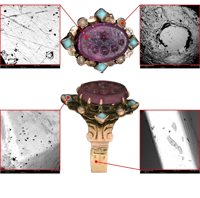
Preservation of Cultural heritage. Research on XIX century jewels for assessing the type manufacturing and the materials used for their production. Courtesy of Francesca Falcone
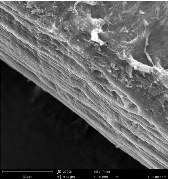
Diagonal cross section of a corneal tissue. Courtesy of Prof. Assunta Pandolfi
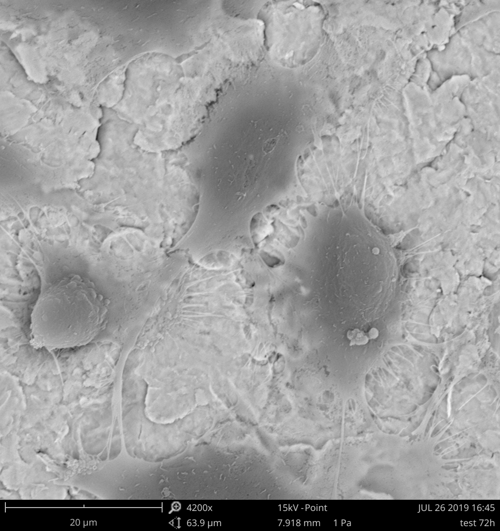
Endothelial cells growing on a Ti alloy substrate. Courtesy of Susi Zara
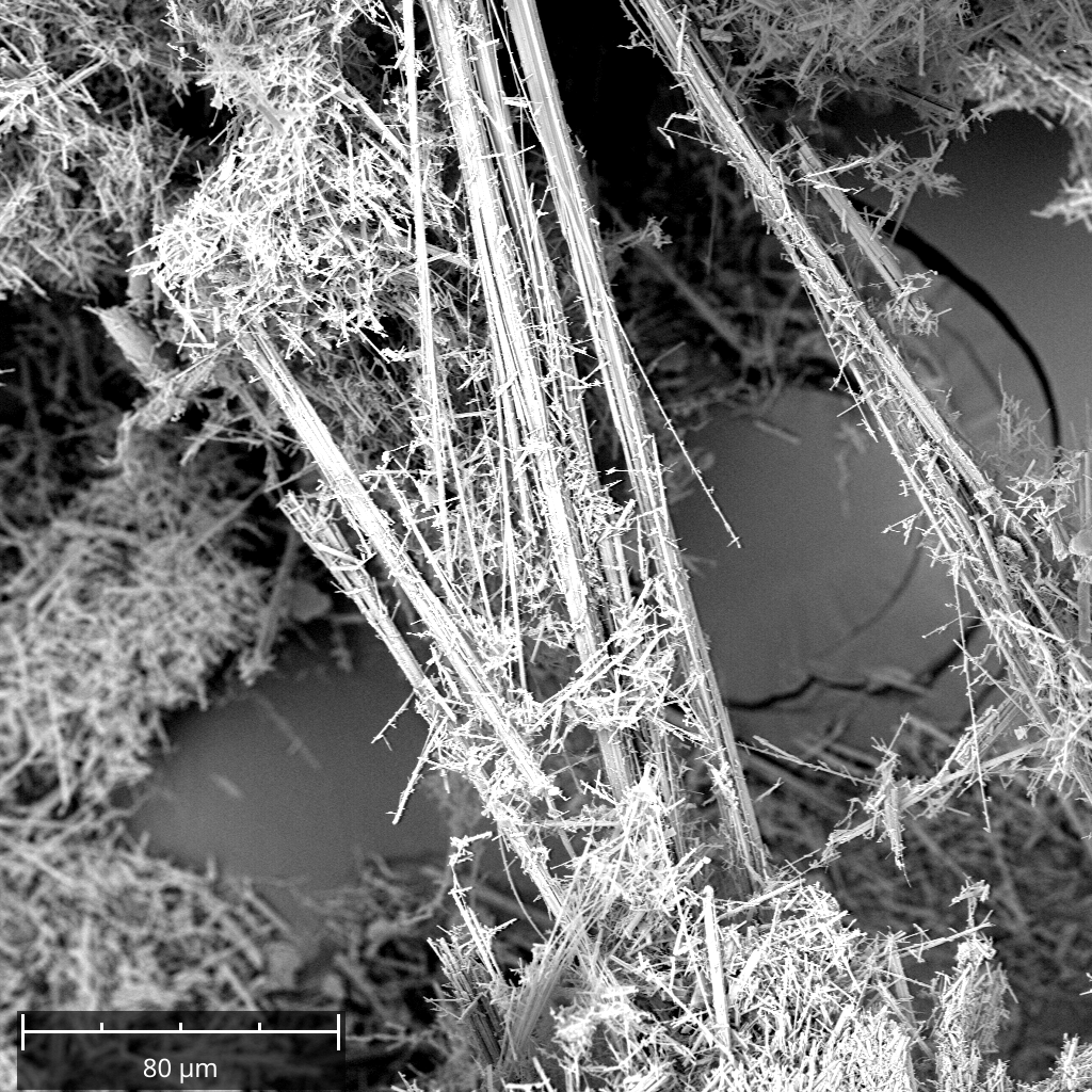
Asbesto fibers. Courtesy of Rosatelli Gianluigi
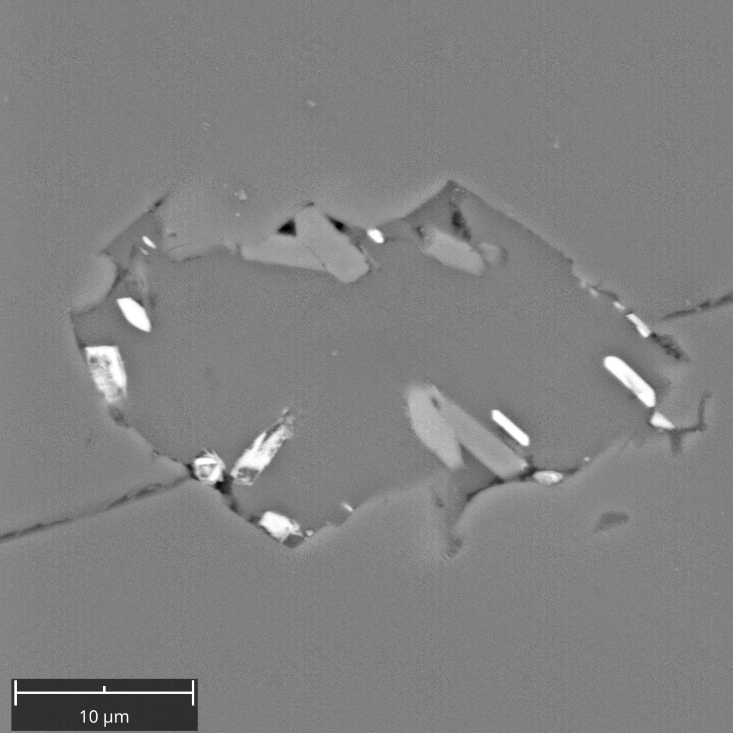
Composite carbonate inclusion in olivine. Carbonate contains microphenocrysts of barite, clinopyroxene and magnetite. Courtesy of Rosatelli Gianluigi
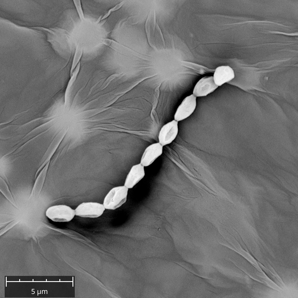
Coliform Bacteria on a composite graphene ultrathin sheet. Courtesy of Maura Mancinelli
PHENOM XL
The Phenom XL scanning electron microscope (SEM) is equipped with CeB6 high luminosity electon source, four-segment backscattered electron detector (BSED), Secondary Electron Detector (SED), computer-controlled motorized X and Y sample stage, fully integrated EDS Silicon Drift Detector (SDD), software package Element Identification (EID) that allows the identification from Boron (5) up to Americium (95).READ MORE
