Transmission Electron Microscopy
The EM facility is a laboratory providing expertise in transmission electron microscopy on a wide range of biological samples, cells in culture and tissue.
TEM allows ultrastructural investigation of organelles and subcellular compartments, and acquisition of high-resolution images. This technique is applied in different research fields such as biology, medicine, and it is also suitable for analysis of inorganic materials.
This Facility offers (on a collaborative basis) to research groups of CAST and of the University G. d’Annunzio, as well as to external collaborators, assistance for the preparation and analysis - both qualitative and quantitative- of a variety of samples. The EM Facility staff can also assist researchers defining up-to-date strategies for preparing and analising specific specimens of interest, and , when applicable, also providing training to external personnel for sample preparation o and use of instrumentations.
INFO Access
Please contact directly the Head of Facility at: simona.boncompagni@unich.it
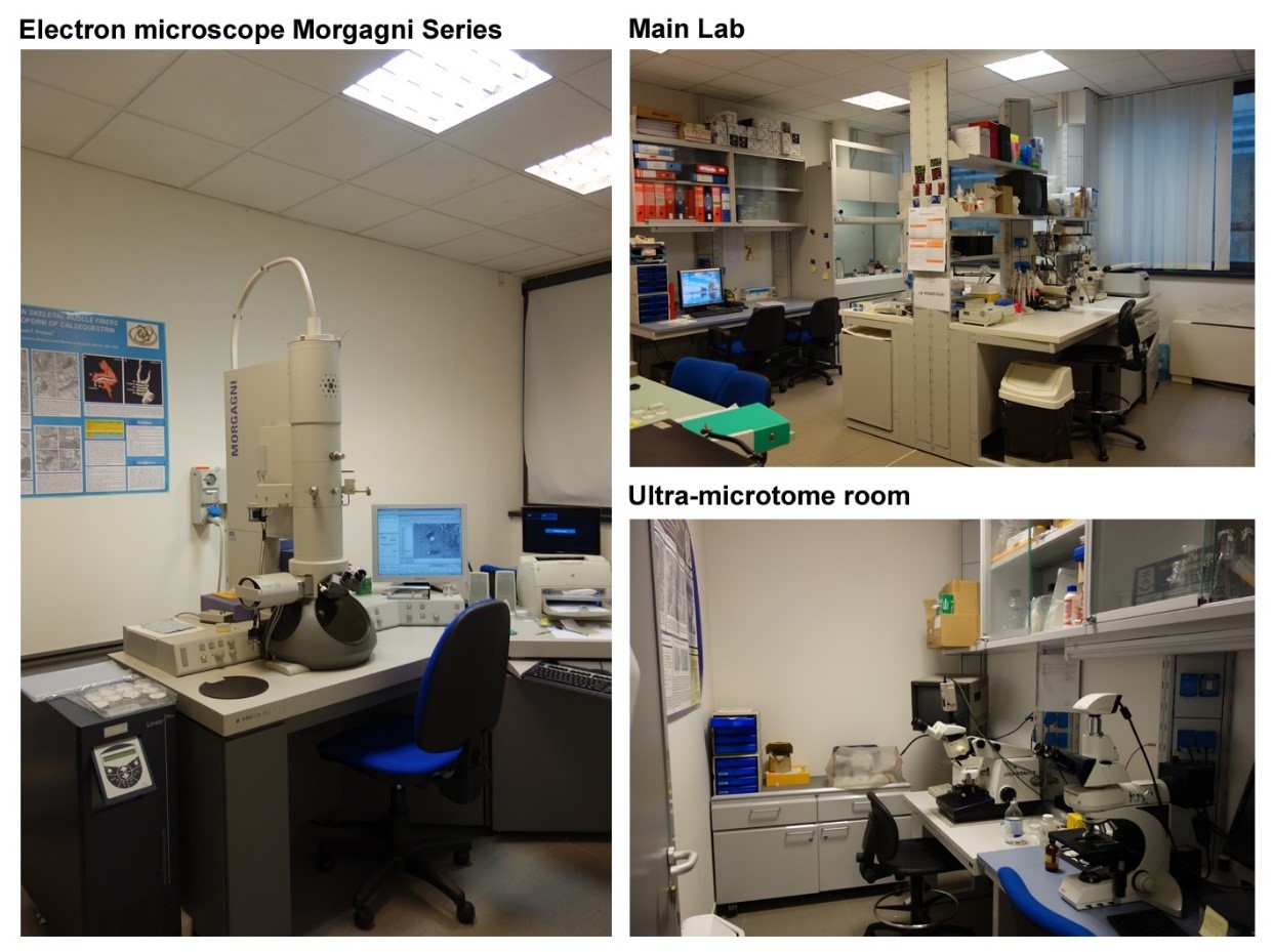
Electron Microscope:
Morgagni Series 268D (FEI Company, Czech Republic) & Megaview III digital camera and Analysis software (Olympus Soft Imaging, Germany)
The Morgagni 268D is a transmission electron microscope. The Electron source is a tungsten tip and the voltage range goes from 40 to 100 kV. It can be used between 1000-180000 magnifications. The microscope is fully controlled by an integrated Dual Pentium PC using Windows 2000 as the main operating system. It is set for fast and easy, high-quality image acquisition and analysis. Image acquisition is done using a side-mounted Olympus Megaview III CCD camera and data analysis is allowed by the Soft Imaging software.READ MORE
Ultra-microtomo:
Leica Ultracut R (Leica Microsystem, Austria)
The Leica Ultracut R produce semi- and ultrathin sections (between 10 nm up to 15 µm thickness) for a wide range of applications. It uses either a glass or diamond knife to cut ultra thin sections of specimens embedded in resins for TEM analysis. Leica Ultracut R/FCS system (Leica Microsystem, Austria) The Ultracut R/FCS is a cryoultramicrotome system specifically designed for routine ultrathin frozen sectioning (-15° to -185°C) for TEM.READ MORE
- Denegri, M., J. E. Rossana Bongianino, F. Lodola, S. Boncompagni, V.C. De Giusti, J. E. Avelino-Cruz, N. Liu, S. Persampieri, A. Curcio, L. Pietrangelo, I. Marty, L. Villani, A. Auricchio, F. Protasi, C. Napolitano, and S. G. Priori. 2014. Single delivery of an adeno-associated viral construct to transfer the CASQ2 gene to knock-in mice affected by Catecholaminergic Polymorphic Ventricular Tachycardia (CPVT) is able to cure the disease from birth to advanced age. Circulation. 129:2673-2681. 10.1161/CIRCULATIONAHA.113.006901.
- Mammucari, C., G. Gherardi, I. Zamparo, A. Raffaello, S. Boncompagni, F. Chemello, S. Cagnin, A. Braga, S. Zanin, G. Pallafacchina, L. Zentilin, M. Sandri, D. De Stefani, F. Protasi, G. Lanfranchi, and R. Rizzuto. 2015. The mitochondrial calcium uniporter controls skeletal muscle trophism in vivo. Cell Reports. 10:1269-1279. 10.1016/j.celrep.2015.01.056
- Boncompagni, S., L. Arthurton, E. Akujuru, T. Pearson, D. Steverding, F. Protasi, and G. Mutungi. 2015. Membrane glucocorticoid receptors are localized in the extracellular matrix and signal through the MAPK pathway in mammalian skeletal muscle fibres. J. Physiol. (London). 593:2679-2692. 10.1113/JP270502
- Pietrangelo, L., A. D'Incecco, A. Ainbinder, A. Michelucci, H. Kern, R.T. Dirksen, S. Boncompagni, and F. Protasi. 2015. Age-dependent uncoupling of mitochondria from Ca2+ release units in skeletal muscle. Oncotarget. 6:35358-35371. 10.18632/oncotarget.6139
- Michelucci, A., A. De Marco, F. Guarnier, F. Protasi and S. Boncompagni. Anti-oxidant treatment (NAC) reduces formation of cores and improves muscle function in RYR1Y522S/WT mice. 2017. Oxid Med Cell Longev. Article ID 6936897. 10.1155/2017/6792694
- Boncompagni, S., A. Michelucci, L. Pietrangelo, R.T. Dirksen, and F. Protasi. 2017. Exercise-dependent formation of new junctions that promote STIM1-Orai1 assembly in skeletal muscle. Scientific Reports. 7(1):14286. 10.1038/s41598-017-14134-0
- Boncompagni, S., A. Michelucci, L. Pietrangelo, R.T. Dirksen, and F. Protasi. 2018. Addendum: Exercise-dependent formation of new junctions that promote STIM1-Orai1 assembly in skeletal muscle. Sci Rep. 8(1):17463. 10.1038/s41598-018-33063-0
- Pietrangelo, L., A. Michelucci, P. Ambrogini, S. Sartini, F.A. Guarnier, A. Fusella, I. Zamparo, C. Mammucari, F. Protasi, and S. Boncompagni. 2018. Muscle activity prevents the uncoupling of mitochondria from Ca(2+) Release Units induced by ageing and disuse. Arch Biochem Biophys. 663:22-33. 10.1016/j.abb.2018.12.017
- Michelucci, A., S. Boncompagni, L. Pietrangelo, M. García-Castañeda, T. Takanoa, S. Malika, R.T. Dirksen and Protasi F. 2019. Transverse Tubule Remodeling Enhances Orai1-dependent Ca2+ Entry in skeletal muscle. Elife. 8:e47576. 10.7554/eLife.47576
- Baraldo, M., A. Geremia, M. Pirazzini, L. Nogara, F. Solagna, C. Türk, H. Nolte, V. Romanello, A. Megighian, S. Boncompagni, M. Kruger, M. Sandri, B. Blaauw. 2020. Skeletal muscle mTORC1 regulates neuromuscular junction stability. J Cachexia Sarcopenia Muscle. 2020.11(1):208-225. 10.1002/jcsm.12496


